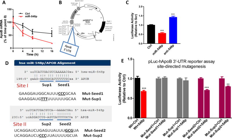
Figure 3. MiR-548p interacts with 3′-UTR of apoB mRNA to promote posttranscriptional degradation.

A. Huh-7 cells were transfected with Ctrl or miR-548p (100 nM). After 24 hours, cells were incubated with actinomycin D (1 μg/ml, from 1mg/ml stock). ApoB mRNA levels at different time points were normlaized using 18S as internal control. The apoB mRNA levels relative to time point 0 were plotted. B. ApoB 3′-UTR was amplified from genomic DNA by PCR and cloned into psiCHECK2 vector at the end of Renilla luciferase cDNA. C. Plasmids (1.5 μg) containing human apoB 3′-UTR were transfected in Huh-7 cells using transfection reagent Turbofect (Life Technologies). After 24 h, Ctrl, miR-548p and anti-548p (100 nM) were introduced into cells. After 48 h, dual luciferase activities were assayed. The data are representative of two independent experiments. D. Two potential targeting sites (Site I and Site II) were predicted by miRanda. ApoB 3′-UTR has complementary bases that could interact with both seed and supplementary sequences. Every seed and supplementary sequence was mutated using site-directed mutagenesis. E. Luciferase reporter plasmids (1.5 μg) containing wild type or different mutant human apoB 3′-UTR were transfected in Huh7 cells. Cells were then transfected with Scr or miR-548p (100 nM). Luciferase activity was measured after 24 h. The data are representative of two independent experiments. p<0.05, **p<0.01, ***p<0.001. Significance was calculated by two-way ANOVA and student’s t-test.
