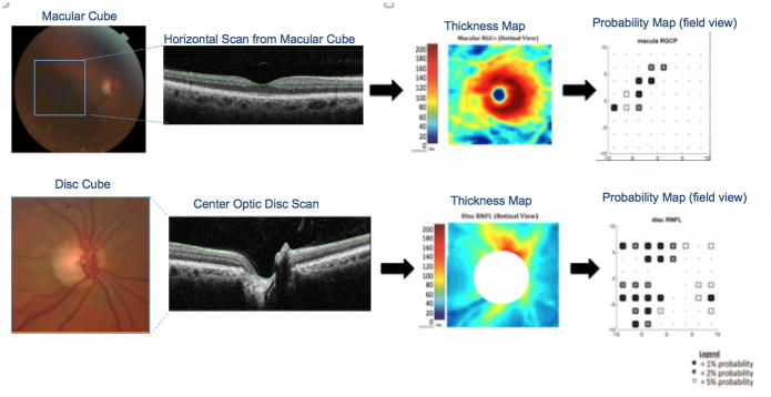
Figure 1.
The combined retinal ganglion cell and inner plexiform layers (RGC+) of the OCT macular scans and the retinal nerve fiber layer (RNFL) of discs scans were segmented using a computer-assisted manual segmentation technique, down-sampled into 64 pixels and converted to a thickness map and then to a probability map.

