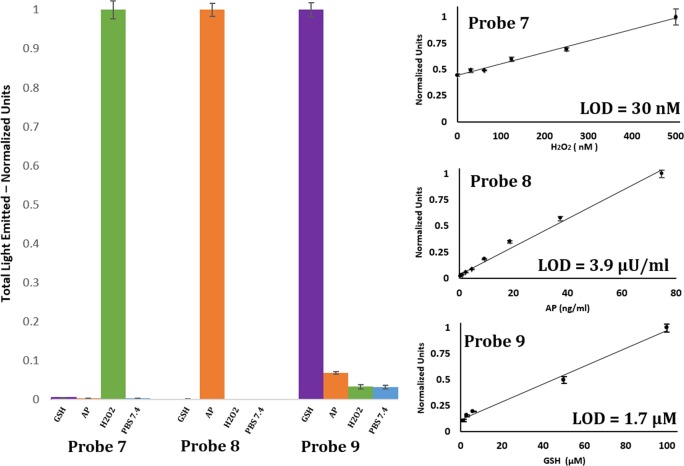
Figure 8.
(Left) Total light emitted from probe 7 [100 μM], probe 8 [10 μM], and probe 9 [10 μM] in the presence of hydrogen peroxide [1 mM, green], alkaline phosphatase [1.5 EU/mL, orange], and glutathione [1 mM, purple]. Measurements were conducted in PBS [100 mM], pH 7.4, with 10% DMSO at room temperature. (Right) Total light emitted from probe 7 [500 μM], probe 8 [500 μM], and probe 9 [10 μM] in PBS [100 mM], pH 7.4 with 10% DMSO, over a period of 1 h over a range of stimulus concentrations. We determined a detection limit (blank + 3 SD) for each probe.

