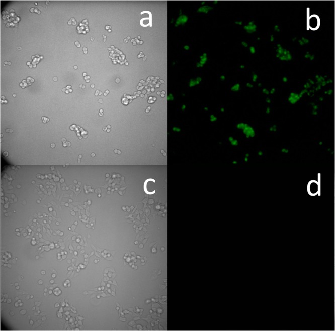
Figure 9.

(a) Transmitted light image and (b) chemiluminescence microscopy image of HEK293-LacZ cells. (c) Transmitted light image and (d) chemiluminescence microscopy image of HEK293-WT cells. Images were obtained following 20 min incubation with cell culture medium containing probe 4 (5 μM). Images were taken using the LV200 Olympus microscope using a 60× objective and 40 s exposure time.
