Figure 2.

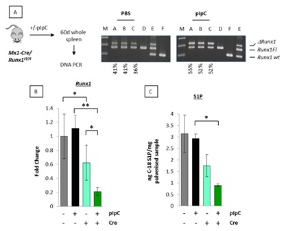
Enforced deletion of Runx1 impairs S1P release in vivo. (A) PCR genotyping of 60‐day‐old splenic tissue DNA from Runx1fl/flMx1Cre+ mice treated with vehicle control (PBS) for background excision or pIpC to excise Runx1. Samples: A–C, 60‐day spleen tissue samples from two groups of three Runx1fl/flMx1Cre+ mice treated with either PBS or pIpC; D–F DNA controls, non‐excised Runx1fl/flMx1Cre− (D), a mixture of partially excised Runx1fl/flMx1Cre+ and Balb/c normal kidney to show all three possible PCR products (E) and Balb/c normal kidney (F). Positions of Floxed (Runx1Fl), deleted (ΔRunx1Fl), and wildtype (WT) Runx1 alleles are as shown. Quantitation to estimate relative excision was performed using image J software. Estimated background and pIpC excision rates are labeled under the blots. (B) qt‐RT‐PCR analysis of Runx1 expression in 60‐day splenic tissue RNA samples from Runx1fl/flMx1Cre+ mice treated with vehicle control or pIpC (dark and light green bars, respectively). Parallel samples were analyzed from Runx1fl/fl mice lacking the Mx1Cre gene to control for PBS and pIpC treatment (black and gray bars, respectively). The data are means ± SD where n = 9 representing three technical replicates of each biological replicate (3). A significant reduction in Runx1 expression was observed between Mx1Cre+ and Mx1Cre− mice indicating background excision (gray vs. light green bar; P = <0.05). Inclusion of pIpC to excise Runx1 generated a further reduction (black vs. green bar; P = <0.01). (C) Remaining splenic tissues were pulverized and analyzed by HPLC mass spectrometry for C18‐S1P. Data are expressed relative to 1 mg of pulverized spleen tissue and represent means ± SD of spleens of three mice for each set of conditions.
