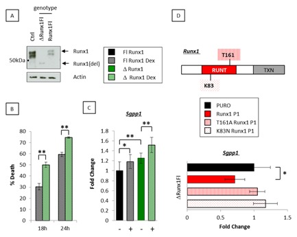
Figure 4.

Enforced deletion of Runx1 promotes dexamethasone‐mediated apoptosis and Sgpp1 transcription. (A) Western blotting analysis as described in Figure 1A to detect the deleted (ΔRunx1Fl) and full length (Runx1Fl) Runx1 proteins from in vitro excised in Runx1fl/flMx1Cre+ 3s B lymphoma cells. (B) Paired cell lines expressing the deleted (ΔRunx1Fl) and full length (Runx1Fl) proteins after in vitro excision of Runx1fl/flMx1Cre+ 3s B lymphoma cells were plated in triplicate in the presence of 1.0 μM dexamethasone and monitored for live/dead counts by trypan blue exclusion. (C) qt‐RT‐PCR analysis of steady state levels of Sgpp1 in ΔRunx1Fl and Runx1Fl 3s cells grown in the presence and absence of 1.0 μM dexamethasone for 6 h. The data are means ± SD where n = 9 representing three technical replicates of each biological replicate (3) from one experiment typical of two. (D) Runx1 schematic showing the mutated residues in the heterodimerization (T161A) and DNA‐binding (K83N) domains. qt‐RT‐PCR analysis of Sgpp1 expression in ΔRunx1Fl 3s cells transfected with full length Runx1, T161A Runx1, or K83N Runx1. Absolute levels of Sgpp1 were compared to control cultures expressing the pBabe Puro vector alone. The data were compiled as described in (C).
