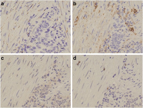
Fig. 1.

Representative photomicrograph of pretreatment biopsy specimens from advanced gastric cancer lesion. a: Immunohistological images of CD42b-positive platelets. Extravasated platelet aggregation (EPA) is mainly seen in the cancer stroma. Cancer-associated fibroblasts (CAFs) with platelet aggregation were observed. b: CAFs in gastric cancer stroma showing D2–40 expression on the membrane, whereas the cancer cells are negative for D2–40 expression. c: SNAIL-positivity expressed in the nuclei of cancer cells. d: Weak expression of forkhead box P3
