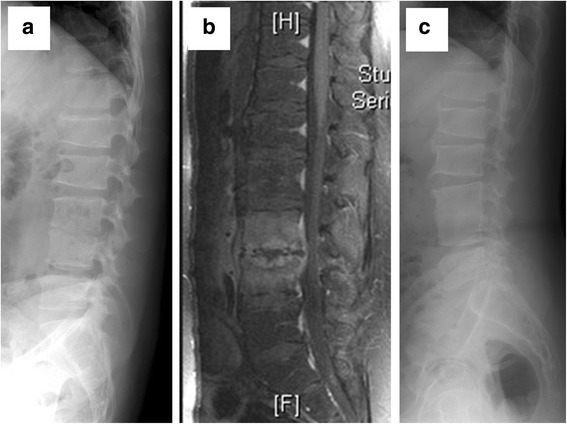
Fig. 2.

Pre- and post-treatment images of a 33 years old male heroin addict in Group 2. a Lateral X-ray view of the spine with pre-treatment kyphosis angle of 3.2°. b Sagittal section of contrast MRI shows disc destruction and end-plate erosion, which indicating pyogenic spondylodiscitis at L3/4. c Lateral X-ray view with post-treatment kyphosis angle of 1.1°, and it revealed bone union after the patient received early surgery using a retroperitoneal approach and L3/4 interbody debridement and fusion, in addition to antibiotics treatment. The patient’s total hospital stay was 25 days, and kyphosis angle correction (1.1–3.2 = −2.1) revealed an improved kyphosis angle and stable spine after surgical correction
