Fig. 6.

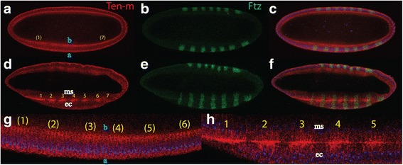
Formation of the Ten-m stripes at gastrulation. a-c confocal picture of a cellularized Drosophila embryo, anterior is to the left and dorsal side up. a Ten-m staining, the protein is detected at the basal (b) as well as apical (a) surface with stronger staining at the basal side where some presumptive stripes emerge, indicated by yellow colors, better seen in a magnification in (g). b identical embryo as in (a), stained for Ftz. The 7 stripes are already established. c merge of (a) and (b). d-f confocal picture of a Drosophila embryo at early gastrulation, anterior is to the left and dorsal side up. d 7 stripes with a width of about 4 cells and a gap of 4 cells have emerged which show strongest accumulation at the interphase between cells at the ectoderm (ec) and the mesoderm (ms), apart from being expressed at the other parts of the cell surface, best seen in a magnification of (d) in (h). e Identical embryos as in (d), Ftz stripes staining both the ectoderm. f merge of (d) and (e)
