Fig. 1.

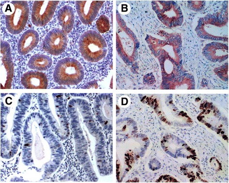
Representative immunohistochemical staining for TUFM and p53 in adenoma and colorectal carcinoma tissue. TUFM staining was positive in adenoma (a) (predominantly localized in the membrane and cytoplasm around the lumen of gland) and in carcinoma (b). Several scattered cells of moderate adenomas displaying p53 staining (c). Carcinoma shows positive staining for p53 (d)
