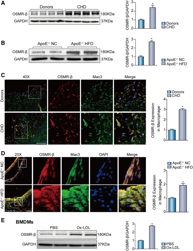
Fig. 1.
Enhanced OSMR-β expression in the atherosclerotic plaques of ApoE−/− mice and patients with CHD. A: The expression of OSMR-β in the coronary arteries of normal donors and patients with CHD (n = 4, *P < 0.05 versus donor). B: The expression of OSMR-β in aortas from ApoE−/− mice fed NC or a HFD (n = 4, *P < 0.05 versus control). C, D: Double immunofluorescence staining for OSMR-β (red) and Mac3 (macrophage, green) in the coronary arteries of normal donors and patients with CHD and in the aortic sinuses from ApoE−/− mice fed NC or a HFD (scale bar = 50 μm). The quantification was carried out by normalizing the fluorescence intensity of the OSMR-β-positive area with the Mac3-positive area in the atherosclerotic plaque. E: OSMR-β expression in BMDMs upon 15 μg/ml Ox-LDL stimulation for 24 h. *P < 0.05 versus control group.

