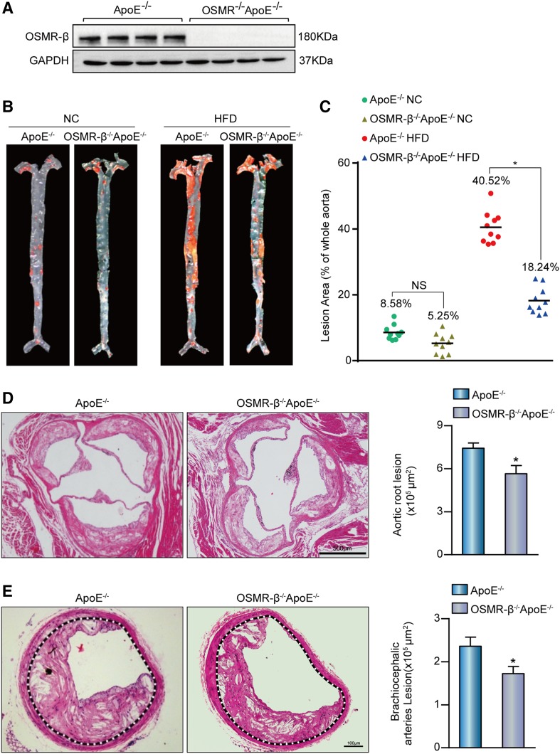
Fig. 2.
Deletion of OSMR-β ameliorated the development of atherosclerosis. A: Genotyping of OSMR-β−/−ApoE−/− mice and ApoE−/− littermates. B, C: En face analysis of aortas from OSMR-β−/−ApoE−/− mice and ApoE−/− littermates fed NC or a HFD; aortas were stained with Oil Red O (n = 10). D, E: Left panel, representative images of the aortic sinus or brachiocephalic arteries from OSMR-β−/−ApoE−/− and ApoE−/− mice stained with H&E. Right panel, quantification of the atherosclerotic lesion area (n = 6). *P < 0.05 compared with ApoE−/−.

