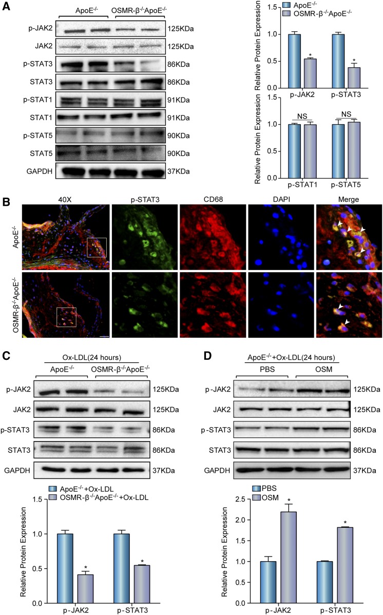
Fig. 5.
OSMR-β deficiency inhibits the activation of JAK2/STAT3 signaling. A: Western blot analysis of the expression of phosphorylated and total JAK2, STAT3, STAT1, and STAT5 in the whole aortas of OSMR-β−/−ApoE−/− and ApoE−/− littermates. Quantitation of relative phosphorylated protein expression after normalization to each total protein expression, respectively (n = 3). B: Immunofluorescence costaining of phosphorylated STAT3 (green) and CD68 (red) in atherosclerotic plaques (scale bar = 50 μm). C: Western blot analysis of phosphorylated and total JAK2 and STAT3 in peritoneal macrophages from OSMR-β−/−ApoE−/− and ApoE−/− littermates upon 15 μg/ml Ox-LDL treatment for 24 h. The results present phosphorylated protein expression compared with the total protein expression, respectively (n = 3). D: The JAK2 and STAT3 expression in macrophages from ApoE−/− mice upon PBS or OSM treatment. *P < 0.05 compared with control group.

