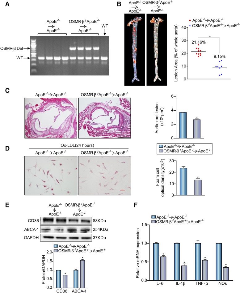
Fig. 6.
The absence of OSMR-β from bone marrow-derived cells attenuates atherogenesis. A: DNA was isolated from whole blood for genotyping to confirm genome reorganization with PCR. B: Left panel, Oil Red O staining for en face analysis of atherosclerotic plaques in aortas from OSMR-β−/−ApoE−/−→ApoE−/− (n = 8) and ApoE−/−→ApoE−/− (n = 10) mice. Right panel, analysis of the atherosclerotic lesion area (n = 10 or 8, each group). C: Left panel, H&E staining of the aortic root from OSMR-β−/−ApoE−/−→ApoE−/− mice and ApoE−/−→ApoE−/− mice. Right panel, quantification of the atherosclerotic lesion area (scale bar = 500 μm, n = 6). D: Oil Red O staining of peritoneal macrophages from OSMR-β−/−ApoE−/−→ApoE−/− mice and ApoE−/−→ApoE−/− mice treated with Ox-LDL. E: Western blot analysis of CD36 and ABCA-1 expression in OSMR-β−/−ApoE−/−→ApoE−/− and ApoE−/−→ApoE−/− mice. F: Real-time PCR analysis of pro-inflammatory cytokine expression in macrophages from OSMR-β−/−ApoE−/−→ApoE−/− mice and ApoE−/−→ApoE−/− mice treated with Ox-LDL (n = 3). *P < 0.05 compared with ApoE−/−→ApoE−/− group.

