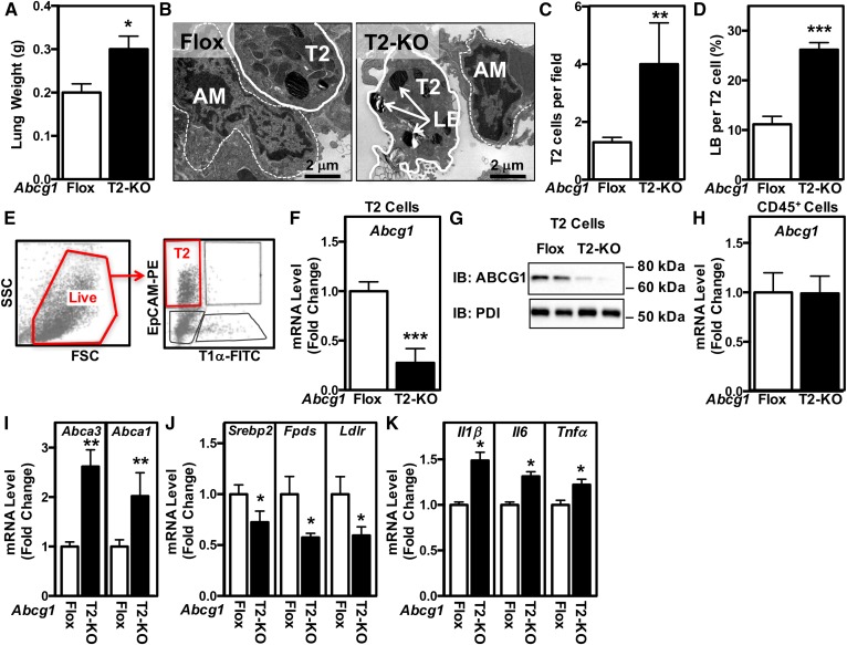
Fig. 2.
Mice with selective deletion of Abcg1 in T2 cells have abnormal surfactant and lamellar body homeostasis. A: The fresh weight of the lungs was increased in Abcg1T2-KO mice. B: Representative electron micrographs from Abcg1flox/flox and Abcg1T2-KO mice (original magnification: 17,400×). Increased T2 cell number (C) and relative area of lamellar bodies within each T2 cell (D). E: Flow cytometry gating strategy to identify T2 cells (defined as EpCAMhiT1α− cells). Single-cell suspensions of negatively selected CD45− cells were stained with fluorophore-conjugated antibodies and analyzed by flow cytometry. Among single cells, the live cells were selected for further analysis to identify T2 cells (EpCAMhiT1α−). F: Abcg1 expression is significantly reduced in EpCAMhiT1α− T2 cells. G: ABCG1 protein is absent from EpCAMhiT1α− T2 cells. H: Abcg1 expression is unchanged in CD45+ cells isolated from Abcg1T2-KO mice. I: Increased Abca3 and Abca1 expression in EpCAMhiT1α− T2 cells. J: Decreased Srebp-2, Fdps, and Ldlr expression in EpCAMhiT1α− T2 cells. K: Increased Il1β, Il6, and Tnfα expression in EpCAMhiT1α− T2 cells. Significance was measured by Student’s t-test. Data are expressed as mean ± SEM. *P < 0.05, **P < 0.01, ***P < 0.001.

