Fig. 5.
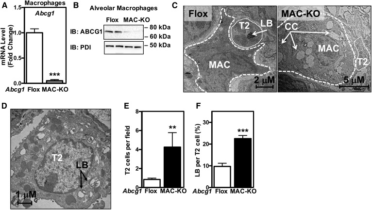
Abcg1−/− macrophages signal to wild-type T2 cells. A, B: ABCG1 is absent in alveolar macrophages isolated from Abcg1MAC-KO mice. A: Abcg1 expression in alveolar macrophages isolated from Abcg1flox/flox and Abcg1MAC-KO mice. B: ABCG1 protein in alveolar macrophages isolated from Abcg1flox/flox and Abcg1MAC-KO mice. C: Representative electron micrographs from Abcg1flox/flox and Abcg1MAC-KO mice (original magnification: 17,400×). D: Electron micrograph of a T2 cell from Abcg1MAC-KO mice (original magnification: 22,600×). Increased T2 cell number (E) and relative area of lamellar bodies within each T2 cell (F) in Abcg1MAC-KO mice. Data are expressed as mean ± SEM (n = 4–6 mice/genotype). AM, alveolar macrophage; CC, cholesterol crystal; LB, lamellar body; T2, T2 cell. Significance was measured by Student’s t-test. **P < 0.01, ***P < 0.001.

