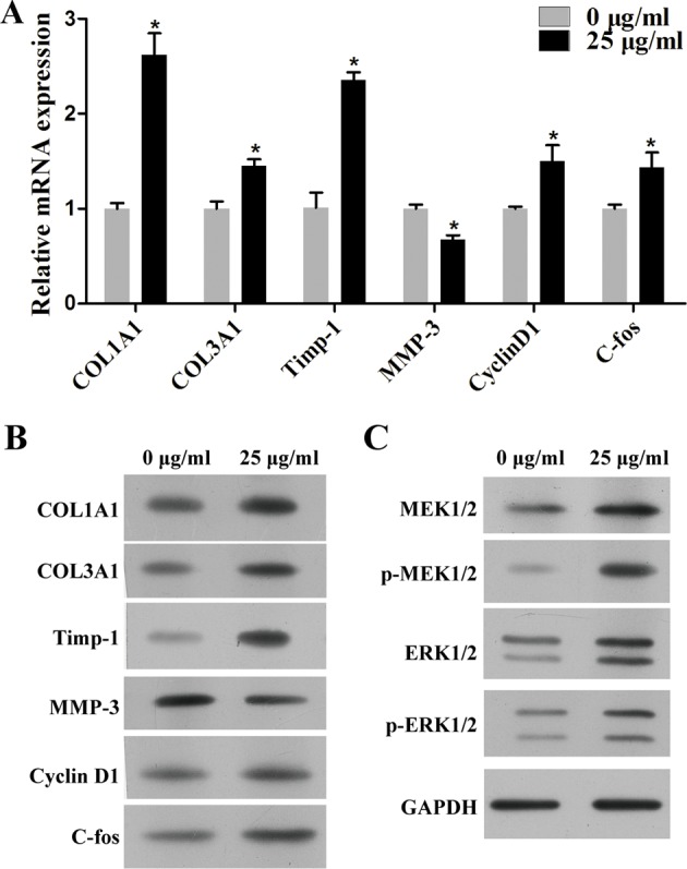
Figure 4. D. genkwa root extractives influence extracellular matrix composition of c-HSF cells througe inucing MEK/ERK signaling pathways.

Mechanism study of D. genkwa in c-HSF cells. (A) qRT-PCR analysis of expression levels of COL1A1, COL3A1, Timp-1, MMP-3, Cyclin D1 and c-fos. The levels of COL1A1, COL3A1, Timp-1, Cyclin D1 and c-fos mRNAs were significantly increased while the level of MMP-3 was down-regulated. *P<0.05 compared with controls. Values are means of three replicates. (B) The protein levels of COL1A1, COL3A1, Timp-1, MMP-3, Cyclin D1 and c-fos in c-HSF cells after treatment with D. genkwa. (C) Western blotting analysis of MEK1/2, p-MEK1/2, ERK1/2 and p-ERK1/2. The protein levels of COL1A1, COL3A1, Timp-1, Cyclin D1, MEK1/2, p-MEK1/2, ERK1/2 and p-ERK1/2 were obviously up-regulated while MMP-3 was down-regulated after the treatment of extracts.
