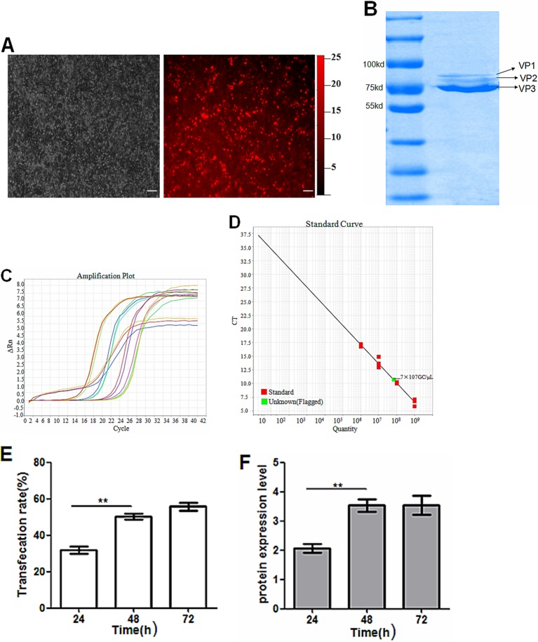
Figure 4. Packaging of viral vector rAAV-iRFP682-TRAIL (bars: 20 μm).
(A) Fluorescence images of HEK-293 cells co-transfected with pAAV-iRFP682-TRAIL and pDG. (B) Purification detection by Coomassie Blue staining method; VP1, VP2, VP3 are the three capsid protein variants of AAV. (C) Amplification plot of TRAIL genes for plasmid standard specimens. (D) Standard curve for plasmids and quantification of viral specimens. (E) pAAV-iRFP682-TRAIL transfection rate after 24, 48 and 72 h. (F) TraiL protein expression levels at 24, 48 and 72 h.

