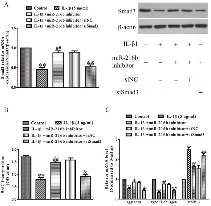
Figure 6. Smad3 was involved in the effects of miR-216b on IL-1β-induced cartilage degradation in SW1353 cells.
SW1353 cells were transfected with either miR-216b inhibitor or si-Smad3, and then treated with IL-1β (5 ng/ml) for 24 h. (A) The mRNA and protein levels of Smad3 were determined by qRT-PCR or Western blot respectively. Smad3 expression was normalized to β-actin. (B) Cell proliferation was assessed by BrdU-ELISA assay. (C) The mRNA levels of aggrecan, type II collagen, and MMP-13 were determined by qRT-PCR. β-Actin was detected as a loading control. All data are presented as mean ± S.E.M., n=4; **P<0.01 versus Control, #P<0.05, ##P<0.01 versus vehicle + IL-1β, &P<0.05, &&P<0.01 versus IL-1β + miR-216b inhibitor.

