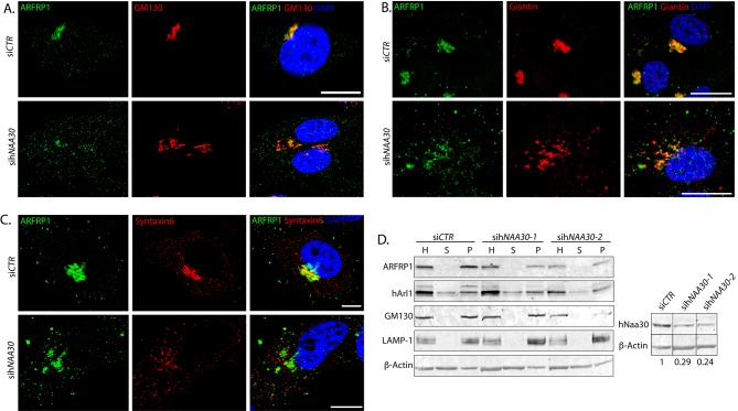
Figure 3. hNaa30 depletion leads to a shift of ARFRP1 from cis-Golgi/TGN-positive compartments to non-Golgi compartments.
Confocal micrographs of Hela cells treated with non-targeting siRNA (siCTR) or sihNAA30 and co-immunolabelled for detection of ARFRP1 and GM130 (A), Giantin (B) or Syntaxin-6 (C). DAPI staining was used to visualize nuclei. White bars indicate 10 μm. (D) Immunoblot of cell lysates from siRNA-treated cells after organelle sedimentation. L, total lysates; P, organelle-enriched pellets; S, supernatant after organelle sedimentation. β-actin was used as a loading control for total cell lysates. GM130 and lysosome-associated membrane glycoprotein 1 (LAMP-1) are used as controls for organelle sedimentation. Knockdown efficiency is shown in the panel to the right, with loading adjusted hNaa30 protein levels given under the hNaa30 immunoblot. hNaa30 blots are taken from the same membrane as rest of the sedimentation and aligned together with loading control for easy visualization.

