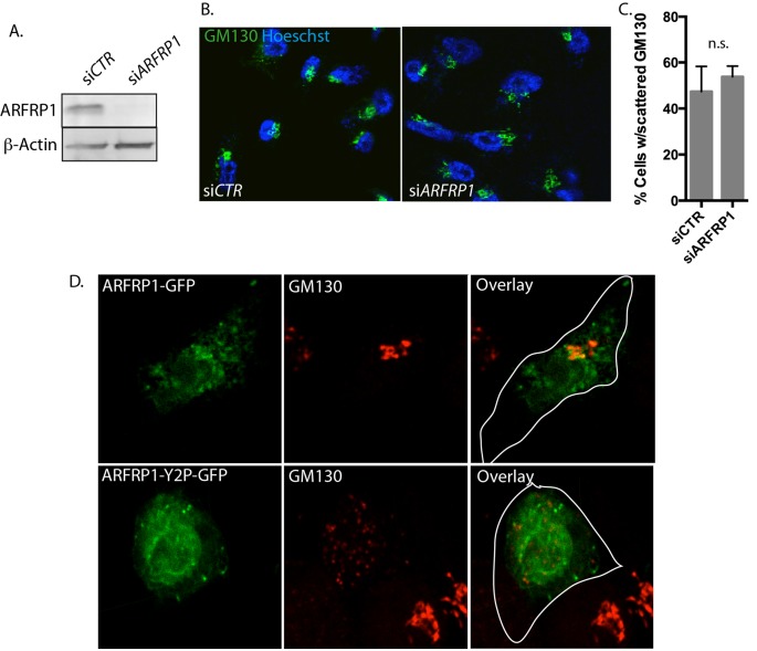
Figure 4. ARFRP1-Y2P-GFP overexpression but not ARFRP1 depletion induces GA fragmentation.
HeLa cells were treated with siCTR or siARFRP1. (A) Protein depletion was verified by immunoblotting. (B) Confocal micrographs of HeLa cells treated as in (A) and immunostained for Golgi protein GM130 (green). Hoescht 33342 was used to visualize the nucleus. (C) Cells displaying scattered Golgi were quantified from at least three independent samples with at least 100 cells counted in each sample, and Student’s t test was used to evaluate differences between conditions (P<0.05). (D) Confocal micrographs of HeLa cells transfected with plasmids encoding ARFRP1-GFP and ARFRP1-Y2P-GFP and immunostained for GM130 (red). Representative images from two independent experiments performed in triplicates are displayed.

