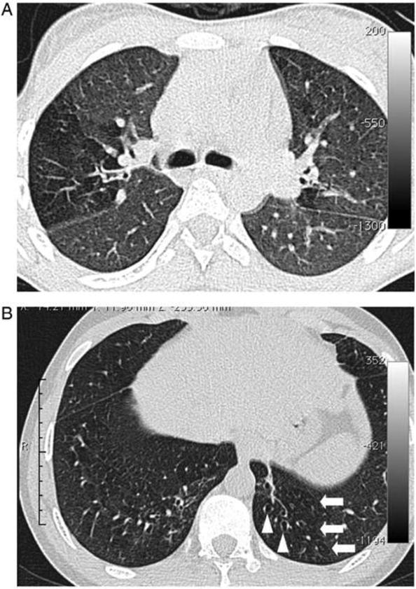
Figure 1.

A, Image section at the level of the carina in a 15-year-old female. There is a clear zone of decreased attenuation in the right upper lobe (and, to a lesser extent, the left lung). In regions of decreased attenuation there is reduction in the caliber of pulmonary vessels; there was no bronchiectasis in this patient. B, Image section in a 19-year-old male through the lower zones demonstrating focal areas of decreased attenuation in both lungs (arrows) and bronchiectasis in the left lower lobe (arrowheads).
