Figure 1.
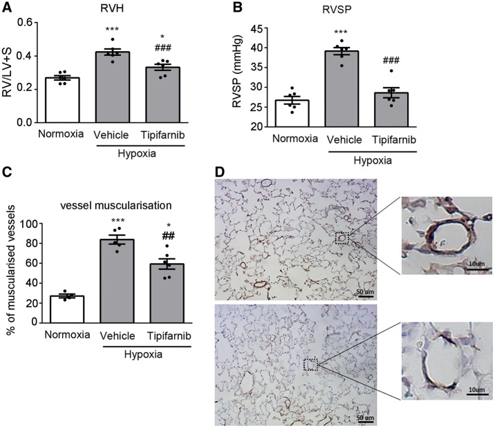
Tipifarnib attenuates development of chronic hypoxia-induced pulmonary hypertension. (A) right ventricular hypertrophy (RVH), (B) Right ventricular systolic pressure (RVSP), (C, D) vessel muscularization in the lungs of normoxic and chronically hypoxic mice treated with vehicle or tipifarnib (100 mg/kg). In (D) top panel shows smooth muscle actin (SMA) staining in lung sections from hypoxic control lungs and bottom panel shows SMA staining in lungs from tipifarnib-treated hypoxic mice. Magnified boxed areas illustrate changes in muscularization of small intrapulmonary arteries. In (A–C) values are means ± SEM of n = 6; *P < 0.05, **P < 0.01, ***P < 0.001 vs. normoxic control; ##P < 0.01, ###P < 0.001 vs. hypoxic control. 1-way ANOVA with Tukey post-test.

