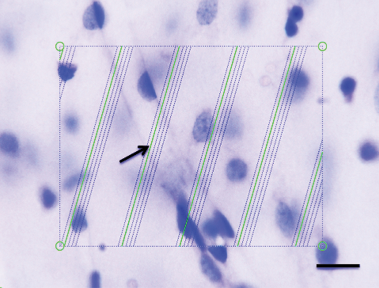
Figure 1.
Estimation of the length of microvessels in a 65-µm-thick vibratome section stained with thionin and visualized on a light microscope with a 60× objective oil immersion lens. Green test lines are superimposed on the live image by newCAST software, and they represent the intersection between isotropic virtual planes and the focal plane. Whenever the microvessels were in focus and virtual planes intersect them, they were counted. One microvessel is intersecting a virtual plane (arrow). Scale bar = 10 µm.

