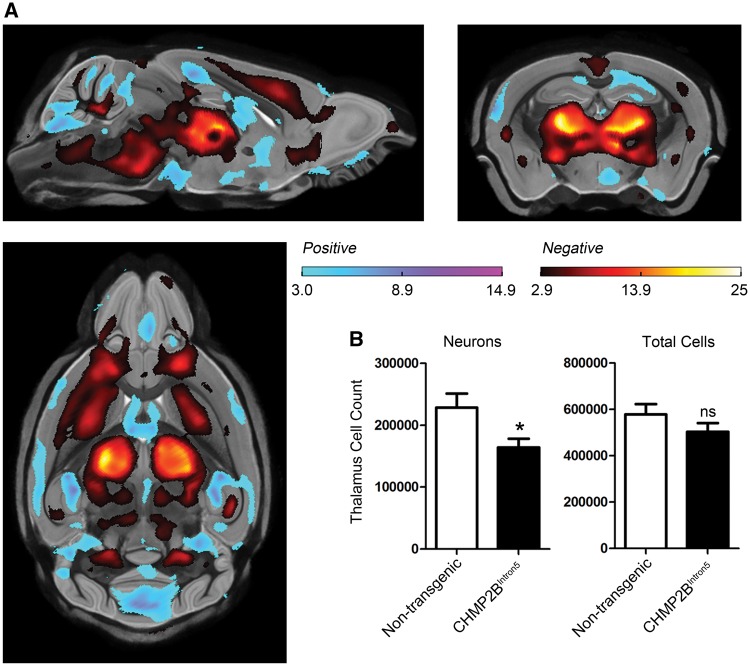
Figure 1.
Volume loss is accompanied by neuronal loss in 18-month-old CHMP2BIntron5 mouse brain. (A) High resolution ex vivo MRI results, showing statistically significant FDR-corrected (q = 0.05) t-statistics overlaid on the group-wise registration average image, revealing local structural differences between the brains of the CHMP2BIntron5 mice (n = 9) relative to non-transgenic controls (n = 10). Regions highlighted in red represent a volume decrease in the CHMP2BIntron5 brains, and regions highlighted in blue represent a volume increase. Significant volume loss can be readily observed in the thalamus, cortex and brain stem of CHMP2BIntron5 brains. Sagittal, coronal and horizontal views are shown. (B) Stereological cell counts reveal a significant decrease in neuron number in 18-month-old CHMP2BIntron5 thalamus (n = 5) when compared with non-transgenic control (n = 5). There is no difference in the total number of cells in the thalamus. *P < 0.05, t-test. Data are shown as mean ± SEM.

