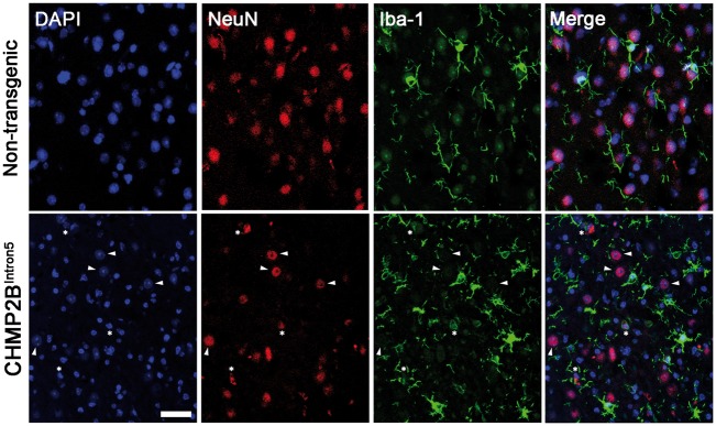
Figure 5.
Microglial engulfment of dying neurons in aged CHMP2BIntron5 mouse brain. Confocal analysis of the association of microglia (Iba1+, green) with neurons (NeuN+, red) in the thalamus of 18-month-old CHMP2BIntron5 mice. DAPI (blue) stains nuclei and is used to identify apoptotic cells (condensed chromatin). Arrowheads indicate normal neuronal nuclei, stars indicate condensed neuronal nuclei characteristic of apoptosis, which are opposed by microglial processes. Scale bar represents 20 µm.

