Figure 7.
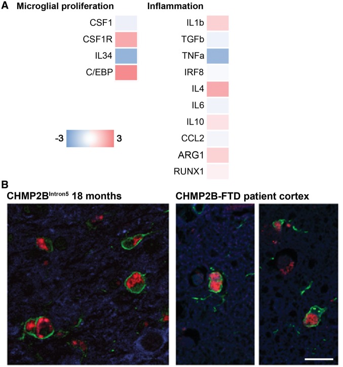
Similar activated microglia profiles in CHMP2B patient brain and 18-month-old CHMP2BIntron5 mouse brain. (A) Analysis of mRNA levels of microglial proliferation and inflammation associated genes in the frontal cortex of CHMP2B-FTD (n = 4) and age-matched controls (n = 5), as performed for 18-month-old CHMP2BIntron5 mice (Fig. 4B). Data are represented with a colour code (blue to red) as fold change of CHMP2B-FTD versus age-matched controls from −3 to 3. (B) Representative images of microglia (Iba1, green) in CHMP2B patient frontal cortex and CHMP2BIntron5 thalamus reveals activated microglia containing characteristic mutant CHMP2B lysosomal storage-like autofluorescent pathology in both patient brain and mouse brain. Autofluorescence is pseudo-coloured in red, neurons (β-III tubulin, blue) are also shown in the left-hand image and nuclei (DAPI, blue) are shown in the right-hand image). Scale bar represents 25 µm.

