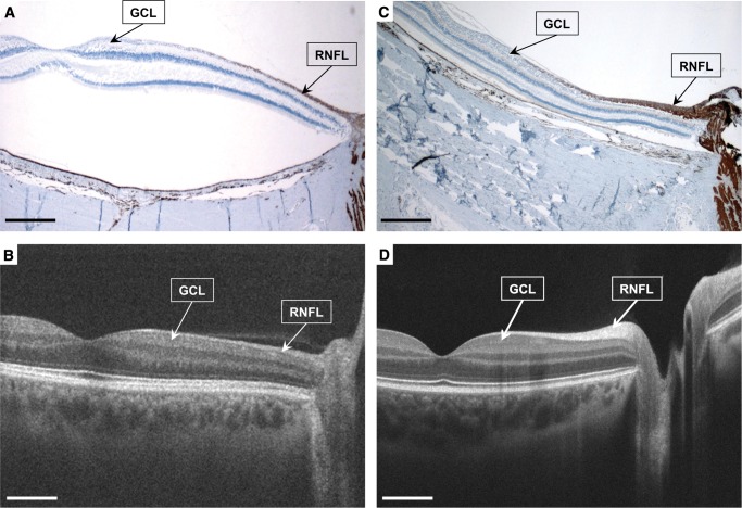
FIGURE 1.
Patient #1 with familial dysautonomia (A) and (B) and aged-matched normal control (C) and (D). Anti-neurofilament immunostained maculopapillary retinal cross-section (2x, (A) and (C)) and matching images from in vivo high definition-optical coherence tomography (B) and (D) show that in the familial dysautonomia patient eye there is marked thinning of the ganglion cell layer ([GCL], mostly midget/parvocellular, P-cells), and their the axons in the retinal nerve fiber layer (RNFL), which occurred primarily in the central retina in the distribution of the maculopapillary bundles (A), retinal detachment is a processing artifact). This was previously documented with the in vivo optical coherence tomography (B). Scale bars, 1 mm.

