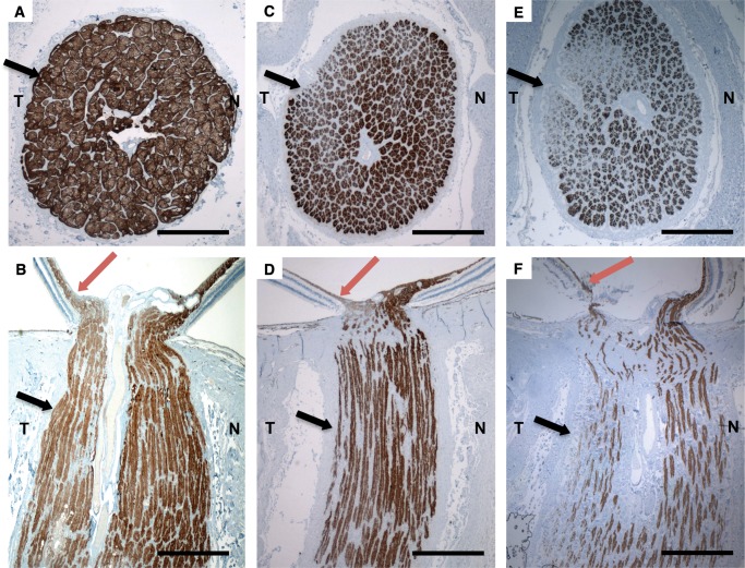
FIGURE 2.
(A–F) Anti-neurofilament-immunostained histopathologic cross and axial sections (2x) of the optic nerve of a 48-year-old normal control (A) and (B) and patients with familial dysautonomia (C–F). Sections from 11-year-old patient #3 (C) and (D) and 61-year-old patient #1 (E) and (F) show significant axonal loss in the optic nerve that is more evident in the temporal optic nerve portion ([T] black arrows), than in the nasal portion [N], as well as thinning in the maculopapillary nerve fiber layer (red arrows). Nerve loss is more prominent in the older patient (Patient #1, (E) and (F)). Scale bars: 1 mm.

