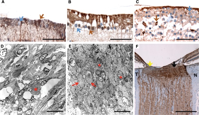
FIGURE 3.
Histopathological cross sections and electron microscopy of the retina and optic nerves of patients with familial dysautonomia. (A–C) Immunohistochemical staining for melanopsin in retinal ganglion cells (40x, scale bars are 50 μm) in Patient #1 (A), Patient #3 (B), and a normal control (C). Macular ganglion cells (stained blue, blue arrow) are markedly reduced in patients with familial dysautonomia (A) and (B) compared to the normal control (C), whereas melanopsin retinal ganglion cells (stained in brown, brown arrow) are preserved in the 3 subjects. Melanopsin retinal ganglion cells are more resistant to mitochondrial stress and are also preserved in Leber hereditary optical atrophy and autosomal dominant optic atrophy (17). (D) and (E) Electron microscopy examination of the prelaminar temporal portion of the optic nerve head in Patient #2 (scale bars: 2 μm). Nerve fibers are arranged in axonal bundles surrounded by astrocytes and a narrow connective tissue space containing capillaries. Low magnification of the optic nerve head shows cells with focal axonal swelling/degeneration (red star). Temporal portion of the optic nerve head pre-laminar region showing nerve bundles (red arrows) with atrophy and mitochondria (red stars). There is axonal swelling with degenerating organelles (lysosomes and mitochondria). Focal myelinated axons were identified beyond the lamina cribrosa area. (F) COX immunostaining (2x, scale bar: 0.5 mm) of the optic nerve of Patient #3 is less intense in the temporal portion ([T], yellow arrow), indicating extensive retinal nerve fiber loss secondary to macular ganglion cell death, compared to more normal staining in the nasal portion ([N], black arrow).

