
Figure 1.
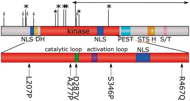
Domain arrangement of DYRK1A with location of diagnostic mutations. The kinase domain is enlarged and the catalytic loop and activation loop are labelled. NLS, nuclear localization signal; DH, DYRK homology box; PEST, proline, glutamic acid, serine, and threonine rich domain; STS, speckle-targeting signal; H, histidine repeat; S/T, serine/threonine repeat. Missense mutations are shown beneath the domain diagram, with position and amino acids; protein truncating variants are show above the diagram (* = stop-gained; lightning-bolt = frameshift; star = splice site; arrow = inversion). Arg437 to a stop codon occurred twice in the dataset.