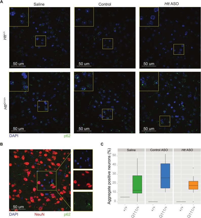
Fig 3. Peripheral Htt silencing does not prevent formation of p62-immunoreactive neuronal intranuclear inclusions in the striatum.
(A) Representative images taken of the dorsolateral striatum show nuclei (DAPI, blue) and autophagy adapter p62 (green) for each genotype and treatment condition. For clarity, staining of neuronal marker NeuN was omitted in (A). However, a neuronal (NeuN) mask was used to selectively quantify p62 aggregates in neurons, therefore a representative image of an HttQ111/+ mouse is shown in (B). Quantification of staining among HttQ111/+ mice (C) reveals no differences in the percent of neurons with intranuclear inclusions between treatment groups.

