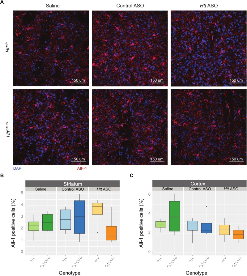
Fig 4. Peripheral Htt ASO treatment does not alter corticostriatal microglia density.
(A) Representative images of AIF-1 (commonly referred to as IBA1) staining taken of the dorsolateral striatum highlight microglia (red) and nuclei (DAPI, blue) in every genotype and treatment condition. No differences were observed in the microglial counts between treatments or genotypes in the dorsolateral striatum (B) or deep cortical layers (C).

