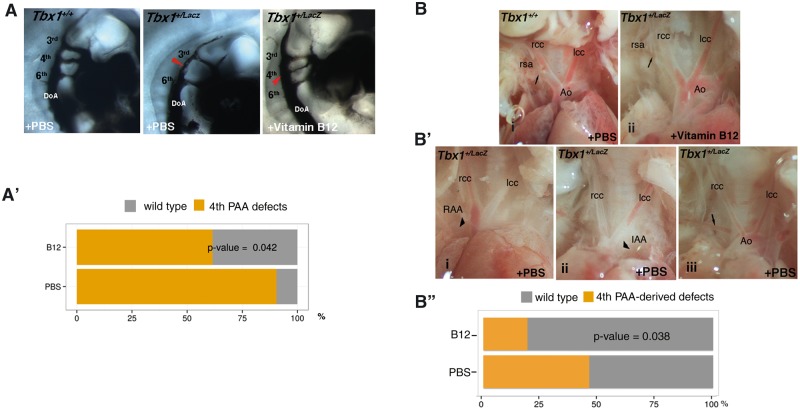
Figure 4.
Vitamin B12 treatment partially rescues the Tbx1 haploinsufficiency phenotype. (A) Pharyngeal arch arteries of E10.5 embryos visualised by ink injection. Lateral view of PBS-treated WT (left panel), PBS-treated Tbx1lacZ/+ (centre), and vitamin B12-treated Tbx1lacZ/+ (right) embryos. (A’) Graphic representation of the percentage of embryos with 4th PAA defects (PBS sample n = 21; vitamin B12-treated sample n = 26). (B) Representative immages of aortic arch and great vessels of E17.5 fetuses. (i) WT pattern; (ii) normal pattern in a vitamin B12-treated Tbx1lacZ/+ fetus; (B’) Examples of aortic arch patterning defects in Tbx1lacZ/+ fetuses: (i) right aortic arch (RAA), the arrowhead indicates the retropositioned arch; (ii) interrupted arch aortic (IAA) type B, the arrowhead indicates the interruption; (iii) retroesophageal right subclavian artery. Arrows indicate the right subclavian artery. Ao: aorta; lcc: left common carotid artery; rcc: right common carotid artery; rsa: right subclavian artery. (B'') Graphic representation of the percentage of fetuses with aortic arch patterning abnormalities derived from 4th PAA defects (PBS sample n = 26; vitamin B12-treated sample n = 26).

