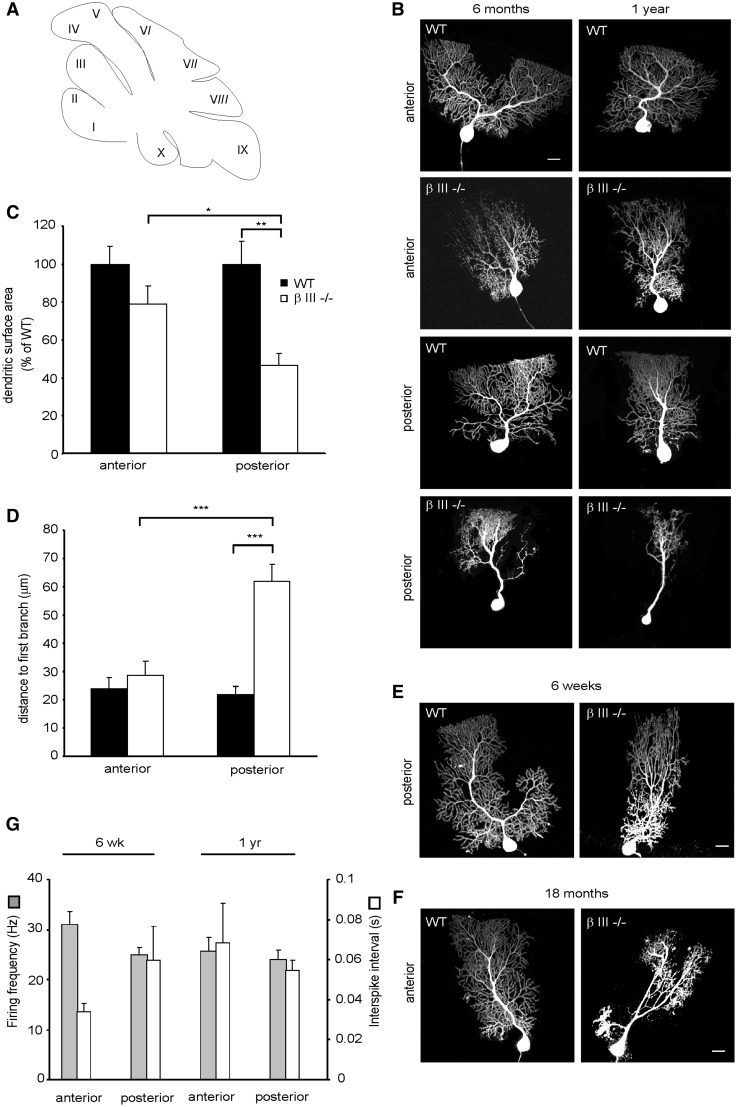
Figure 7.
Proximal dendrites of posterior βIII-/- Purkinje cells most vulnerable to dendritic degeneration. (A) Schematic of sagittal section of cerebellar lobules. (B) Representative confocal images of Purkinje cells filled with Alexa Fluor 568 from anterior (II –V) and posterior (VIII-X) lobules of 6-month and 1-year-old WT and βIII-/- animals. (C) Quantification of dendritic surface area of individual Purkinje cells from 6-month old WT and βIII-/- mice. N = 3, n = 4 (WT), 6 (βIII-/-) for each region. (D) Quantification of distance from Purkinje cell soma to first dendritic branch point in animals > 6-months of age. N = 9, n = 14 (WT), 18 (βIII-/-). (E) Representative confocal images of Purkinje cells filled with Alexa Fluor 568 from posterior (VIII-X) lobules of 6-week old WT and βIII-/- animals. (F) Representative confocal images of Purkinje cells filled with Alexa Fluor 568 from anterior lobules (II –V) of 18-month- old WT and βIII-/- animals. (G) Spontaneous firing frequency and interspike interval of Purkinje cells from young [N= 4, n = 12 (anterior), 19 (posterior)] and old βIII-/- animals [N = 9, n =28 (anterior), 30 (posterior)]. All data are means ± SEM. Bar, 20 μm.

