Figure 5.

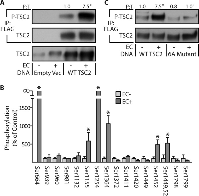
Identification of the eccentric contraction-regulated phosphorylation sites on TSC2. TAs from wild-type male C57 mice were transfected with 30 μg of either the control DNA (empty vector (Empty Vec)), FLAG-tagged WT TSC2, or a FLAG-tagged phosphodefective mutant of TSC2 in which the eccentric contraction-regulated phosphorylation sites were mutated to alanines (6A Mutant). Seven days later, the TAs were stimulated with a bout of eccentric contractions (EC+) or the control condition (EC−) and collected at 1 h after stimulation. A and C, whole homogenates and FLAG immunoprecipitations (IP:FLAG) were subjected to Western blotting analysis for the indicated proteins. B, FLAG immunoprecipitates of WT TSC2 were isolated and then subjected to in-gel trypsin digestion followed by LC/MS/MS to identify and quantify sites of phosphorylation. The values above the blots represent the phosphorylated (P) to total protein ratio (P:T) for each group. All values are presented as means (±S.E. in graph, n = 4–8/group). Note: the values presented for WT TSC2 in A and C are from the same data set. Symbols indicate significant difference (p ≤ 0.05) from DNA matched control (EC−) (*) and stimulation-matched WT TSC2 (•).
