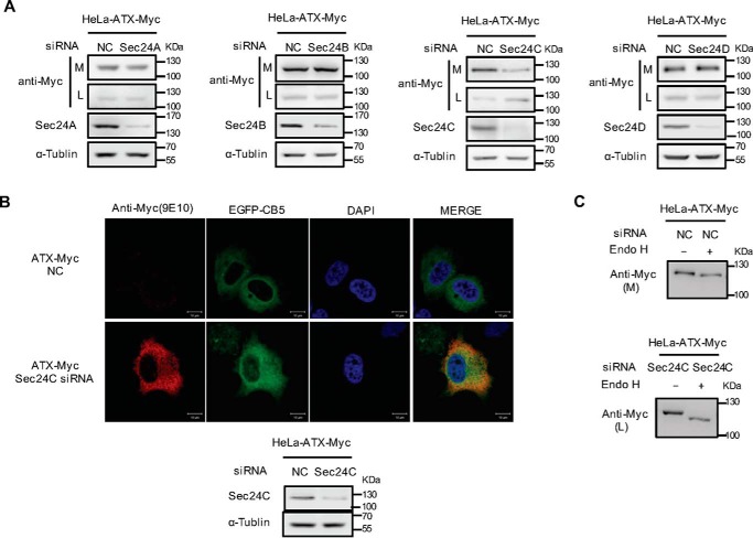
Figure 3.
Identification of Sec24 isoform involved in ER export of ATX. A, HeLa cells were transfected with pcDNA3-ATX-Myc. Twenty four hours after transfection, cells were transfected with nonspecific control siRNA (NC) or siRNA against the indicated Sec24 isoform. Forty eight hours after siRNA transfection, the ATX-Myc protein levels in cell lysates (L) and culture medium (M) were detected by immunoblotting with anti-Myc antibody. B, HeLa cells were co-transfected with pcDNA3-ATX-Myc and pEGFP-N1-CB5 and then treated with nonspecific control siRNA or Sec24C siRNA for 48 h. ATX-Myc was visualized by confocal microscopy with anti-Myc (9E10) monoclonal antibody (red). Endoplasmic reticulum was labeled by EGFP-CB5 (green). Nuclei were counterstained with DAPI (blue). C, HeLa cells were transfected with pcDNA3-ATX-Myc. Twenty four hours after transfection, cells were transfected with nonspecific control siRNA or Sec24C siRNA for 48 h. The concentrated (∼30-fold) serum-free conditional culture medium of the control siRNA-treated cells and the lysate of Sec24C siRNA-treated cells were treated with Endo H and then analyzed by Western blotting with anti-Myc antibody. Data are representative of three independent experiments.

