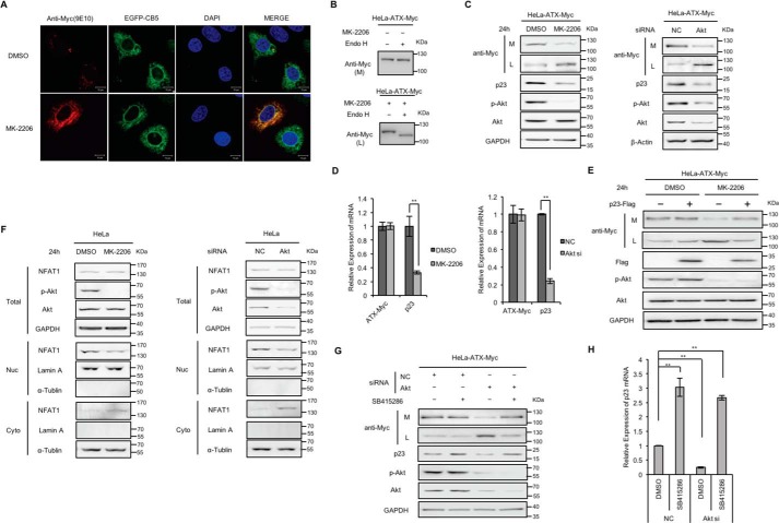
Figure 5.
Effects of AKT inhibitor treatment and AKT knockdown on ATX secretion. A, HeLa cells were co-transfected with pcDNA3-ATX-Myc and pEGFP-N1-CB5 and then treated with or without AKT inhibitor MK-2206 (5 μm) for 24 h. ATX-Myc was visualized by confocal microscopy with anti-Myc (9E10) monoclonal antibody (red). Endoplasmic reticulum was labeled by EGFP-CB5 (green). Nuclei were counterstained with DAPI (blue). B, HeLa cells were transfected with pcDNA3-ATX-Myc and then treated with or without AKT inhibitor MK-2206 (5 μm) for 24 h. The concentrated (∼30-fold) serum-free conditional culture medium of the control cells and the lysate of AKT inhibitor-treated cells were treated with Endo H and then analyzed by Western blotting with anti-Myc antibody. C, HeLa cells with exogenous expression of ATX-Myc were treated with MK-2206 (5 μm) for 24 h or treated with siRNA against AKT for 48 h. Then ATX-Myc protein levels in cell lysates (L) and culture medium (M) were detected by immunoblotting with anti-Myc antibody. p23, AKT, and phosphorylated AKT (p-AKT) levels in cell lysates were detected by immunoblotting. D, HeLa cells with exogenous expression of ATX-Myc were treated with MK-2206 (5 μm) for 24 h or treated with siRNA against AKT for 48 h. ATX-Myc and p23 mRNA levels were detected by quantitative reverse transcription PCR (qRT-PCR). E, HeLa cells with exogenous expression of ATX-Myc and FLAG-p23 were treated with or without AKT inhibitor MK-2206 (5 μm) for 24 h. ATX-Myc protein levels in cell lysates and culture medium were detected by immunoblotting with anti-Myc antibody. FLAG-p23 levels in cell lysates were detected by immunoblotting with anti-FLAG antibody. F, HeLa cells were treated with MK-2206 (5 μm) for 24 h or treated with siRNA against AKT for 48 h. Then, NFAT1 levels in nuclear fraction (Nuc), cytoplasmic fraction (Cyto), and total cell lysates (Total) were detected by Western blotting. Lamin A and α-tubulin were used as the nuclear and cytoplasm markers, as indicated. G and H, HeLa cells with exogenous expression of ATX-Myc were transfected with nonspecific control siRNA (NC) or siRNA against AKT. Thirty two hours after transfection, cells were treated with or without GSK3β inhibitor SB415286 (25 μm) for 16 h. Then, ATX-Myc protein levels in cell lysates and culture medium, and p23 levels in cell lysates were detected by immunoblotting (G). p23 mRNA levels were detected by qRT-PCR (H). The p value was derived from analysis of variance. **, p < 0.005. Data are representative of three independent experiments.

