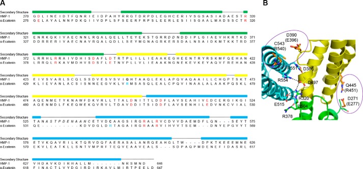
Figure 3.
Sequence alignment between HMP-1 M and αE-catenin M. A, structure-based sequence alignment of the HMP-1 M domain with mouse αE-catenin M domain. Conserved residues involved in forming salt bridges are labeled in red; the italicized residues are not visible in the structure. The colored rectangular box on top of the sequence represents the helical region. B, residues in the HMP-1 M domain structure homologous to those from αE-catenin that form the six salt bridges that stabilize the αE-catenin M domain. Polar interactions are shown as dashed lines in the four conserved salt bridges. Non-conserved residues are circled and corresponding residues of αE-catenin are colored orange and labeled in parentheses.

