Figure 5.
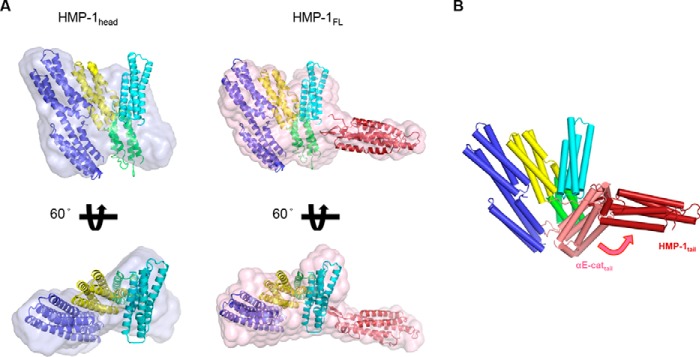
Model of full-length HMP-1. A, a HMP-1 full-length model constructed within an averaged and filtered envelope from SAXS data analysis is shown together with the SAXS envelope of the HMP-1 head in two different orientations. The HMP-1 M domain and crystal structures of the N and C domains of αN-catenin (PDB codes 4P9T and 4K1O) were fitted within the molecular envelope using the head domain of αE-catenin as a guide (PDB code 4IGG). The N and C domains are colored blue and red, respectively. B, structural alignment of the HMP-1 full-length model with one protomer of the dimeric structure of αE-catenin (PDB code 4IGG). The aligned head domain of αE-catenin is omitted and the differently positioned tail domain of αE-catenin is shown and colored salmon.

