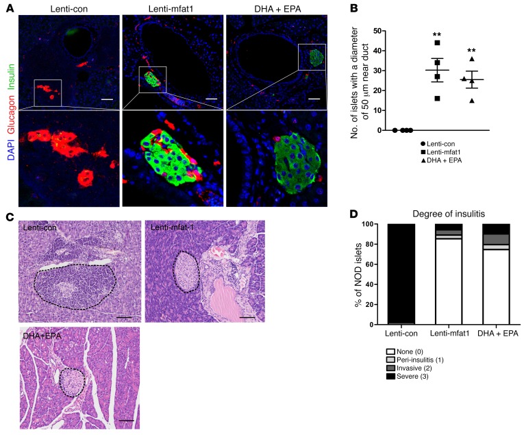
Figure 6. ω-3 PUFAs have a therapeutic effect on immune infiltration in diabetic NOD mice.
Confocal images (A) and quantification (B) of islets with a diameter of 50 μm that appeared adjacent to pancreatic ducts in diabetic NOD mice after lentivirus treatment and DHA plus EPA dietary intervention for 9 weeks (n = 4/group). Scale bars: 50 μm. Original magnification: ×400. **P < 0.01 versus the lenti-con group (Student’s t test). Values represent the mean ± SEM. (C) H&E-stained sections of islets from pancreatic tissue obtained from diabetic NOD mice after lentivirus treatment and DHA plus EPA dietary intervention for 9 weeks. Scale bars: 50 μm. Images are representative of 3 biological replicates. (D) Quantification of the incidence of insulitis in diabetic NOD mice after lentivirus injection or DHA plus EPA dietary intervention for 9 weeks (n = 4/group). Islets were sorted into 4 categories on the basis of the relative degree of immune infiltration: no insulitis (0), peri-insulitis (1), invasive insulitis (2), and severe insulitis (3). The differences in the incidence of no insulitis or severe insulitis between the lenti-con and lenti-mfat-1 groups (P < 0.0001) and between the DHA plus EPA and lenti-con groups (P < 0.0001) were significant. Statistical significance was determined by Pearson’s χ2 test.

