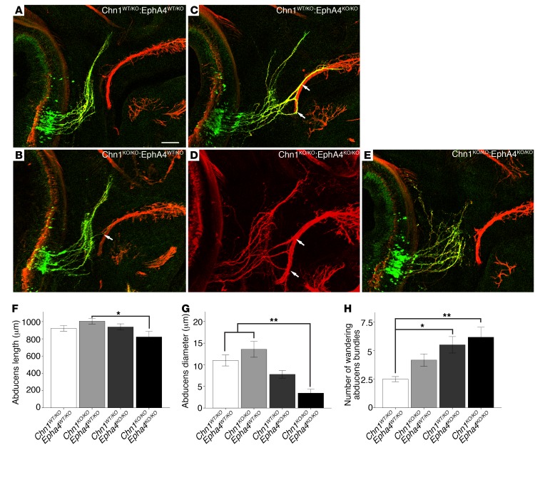
Figure 4. Chn1KO/KO Epha4KO/KO embryos display reduced abducens nerve diameter and enhanced nerve wandering.
(A–E) Whole mount staining of E11.5 abducens nerves in Chn1WT/KO Epha4WT/KO (A; n = 3 embryos), Chn1KO/KO Epha4WT/KO (B; n = 4 embryos), Chn1WT/KO Epha4KO/KO (C; n = 4 embryos), and Chn1KO/KO Epha4KO/KO (D and E; n = 5 embryos) embryos. Arrows, abducens nerve fasciculation with buccal branch of facial nerve; 3/5 Chn1KO/KO Epha4KO/KO E11.5 embryos have abducens phenotypes similar to that in D and 2/5 are similar to that in E. Scale bar: 100 μm. (F–H) Abducens nerve length from hindbrain exit to most distal projection (F); abducens diameter at orbit (G); and number of wandering abducens nerve bundles (H). *P < 0.05, **P < 0.01, 1-way ANOVA with Tukey’s test; data represent mean ± SEM.

