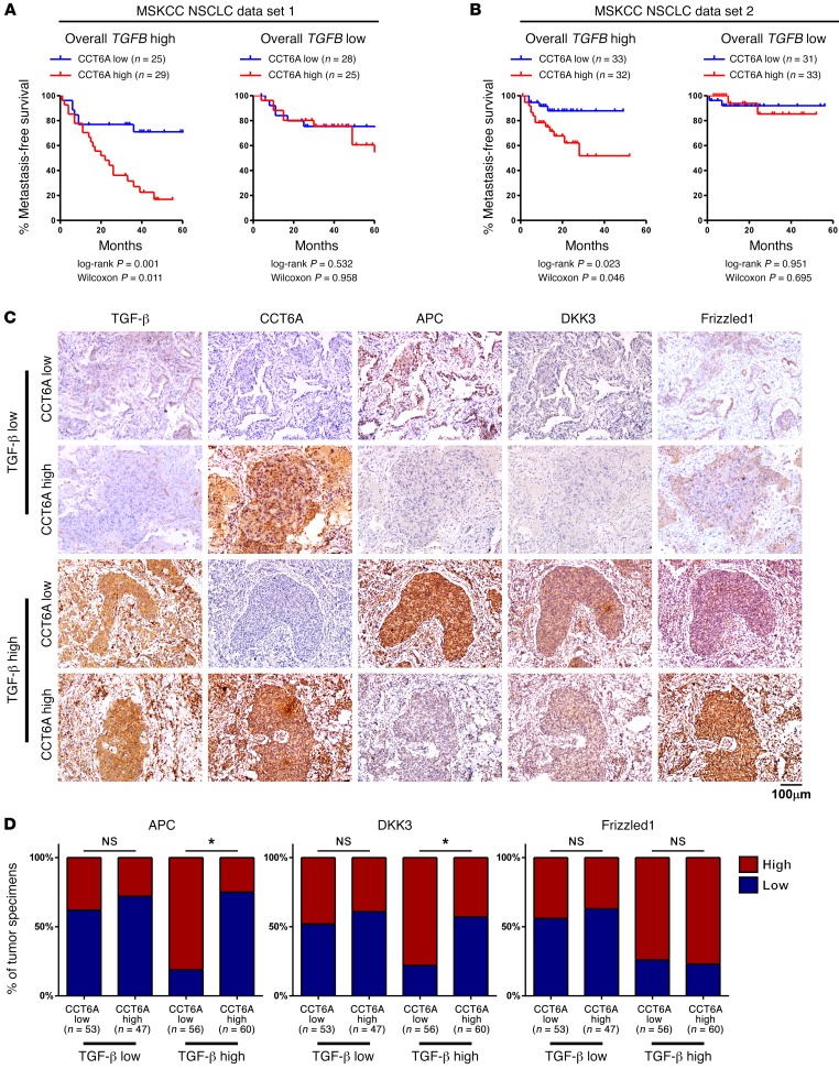
Figure 7. High CCT6A levels are associated with inhibition of the SMAD2-mediated transcriptional program in clinical NSCLC specimens.
(A and B) Kaplan-Meier survival analysis of data collected from MSKCC NSCLC data sets 1 and 2 indicated that CCT6A expression levels were positively correlated with metastasis in patients with high TGF-β expression levels. (C and D) Representative images (C) of immunohistochemical staining for TGF-β, CCT6A, APC, DKK3, and Frizzled1 and statistical analysis across 216 NSCLC specimens (D) show that CCT6A expression was negatively associated with expression of APC and DKK3 only in specimens with high TGF-β levels and that Frizzled1 expression was not correlated with CCT6A expression, regardless of TGF-β levels. “High” and “low” expression levels of each protein were stratified by the median optical density (OD) of staining in all specimens. Scale bar: 100 μm. *P < 0.05, by 1-way ANOVA.

