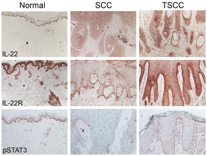
Fig. 4.
IL-22 cytokine, receptor, and downstream signaling is enhanced in TSCC. Immunohistochemistry performed on normal, SCC and TSCC shows enhanced expression of IL-22, IL-22R, and pSTAT3. Frozen tissue sections of normal skin and SCCs and TSCCs were stained with the following antibodies: IL-22 (R&D systems), IL-22 Receptor (Prosci), and pSTAT3 (Santa Cruz Biotechnology). Biotin-labeled horse anti-mouse antibody (Vector Laboratories) was then amplified with avidin-biotin complex (Vector Laboratories) and developed with a chromogen 3-amino-9-ethylcarbazole (Sigma Aldrich). Counterstaining was then carried out with light green (Sigma-Aldrich). With each immunohistochemistry experiment the appropriate isotype controls were performed. (10× magnification). (Adapted from Zhang S, Fujita H, Mitsui H, et al. Increased Tc22 and Treg/CD8 ratio contribute to aggressive growth of transplant associated squamous cell carcinoma. PLoS One 2013;8(5):e62154; with permission.)

