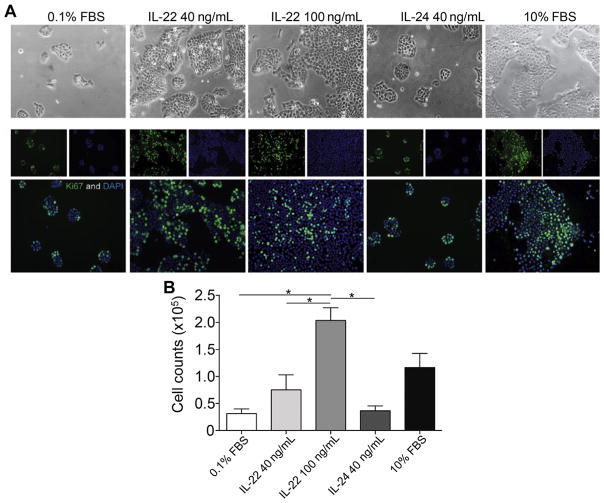
Fig. 5.
IL-22 treatment enhances human cutaneous SCC proliferation. (A) A431 cells were cultured in 0.1% fetal bovine serum (FBS) serum starvation conditions with or without the indicated cytokines for 72 hours. Cells cultured in the presence of IL-22 cytokine or 10% FBS showed increased colony formation compared with those grown under serum starvation conditions or treated with IL-24. The phase images (below) are representative immunofluorescence images staining for the Ki-67 proliferation marker (green) and nuclear 4,6-diamidino-2-phenylindole (DAPI) (blue). (B) Cell counts were performed after 72 hours in the indicated conditions and show enhanced cell numbers after treatment with control 10% FBS and IL-22 conditions (1-way analysis of variance [ANOVA], P<.001). *p< 0.05, determined by one-way ANOVA. (From Zhang S, Fujita H, Mitsui H, et al. Increased Tc22 and Treg/CD8 ratio contribute to aggressive growth of transplant associated squamous cell carcinoma. PLoS One 2013;8(5):e62154; with permission.)

