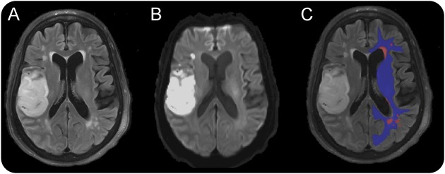
Figure 1. Image processing steps (here shown on 3T MRI of an acute stroke patient) of deriving NAWM masks in the contralesional hemisphere.
Sample FLAIR image (A), acute DWI scan depicting ischemic stroke in the right frontal operculum (B), and representative WMH mask (red) and NAWM mask (blue) overlaid on FLAIR image (C). DWI = diffusion-weighted imaging; FLAIR = fluid-attenuated inversion recovery; NAWM = normal-appearing white matter; WM = white matter; WMH = white matter hyperintensity.

