Figure 1.
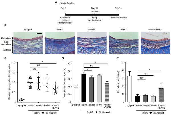
Combined treatment with relaxin and LOX inhibition attenuates established fibrosis in the OTT model. Tracheal segments from donor mice were transplanted orthotopically into major histocompatibility complex (MHC)-matched (syngraft) or mismatched (allograft) recipient mice on day 0. (A) Non-immunosuppressed OTT mice were subjected to treatment at day 21, when fibrosis is well-established, for 14 days. Mice were treated with recombinant human relaxin-2 or saline, with or without the LOX inhibitor, 0.2% β-Aminopropionitrile (BAPN). (B) Representative images of Masson trichrome staining of tracheal cross-sections, in which collagen was stained in blue. Scale bar = 50 μm. (C) Hydroxyproline concentration in tracheal hydrolysates relative to controls was measured to assess the amount of collagen; Mean ± SD. (D) Analysis of collagen density in trichrome stained sections measured by the ratio of the blue area to the area between the subepithelium and cartilage with Image J; Mean ± SEM. (E) Measurement of epithelial thickness in tracheal cross-sections from different groups; Mean ± SEM. *P<0.05. For B, D, E, n=3-10 per group with at least 10 tissue sections per animal.

