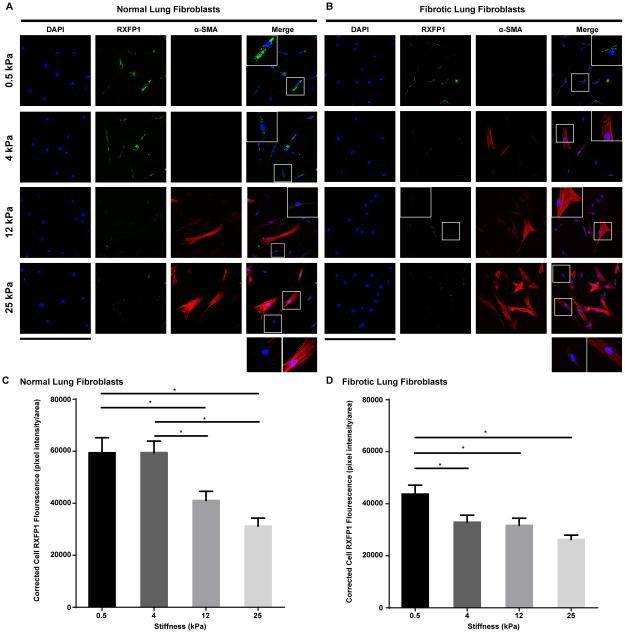
Figure 4.
Expression of relaxin receptor 1 (RXFP1) on fibroblasts. (A, B) Immunofluorescent staining of RXFP1 on normal (A) and fibrotic (B) lung fibroblasts cultured on matrices with Young’s elastic moduli of 0.5-25 kilopascal (kPa). Fibroblast to myofibroblast differentiation was assessed by α-smooth muscle actin (α-SMA) expression; scale bar =424 μm; inserts are 4X magnification of selected area. (C, D) Corrected total cell fluorescence of normal (C) and fibrotic (D) lung fibroblasts cultured on matrices with different elastic moduli (measured by Image J). Mean ± SEM. n=40-50 per group, *P<0.05.

