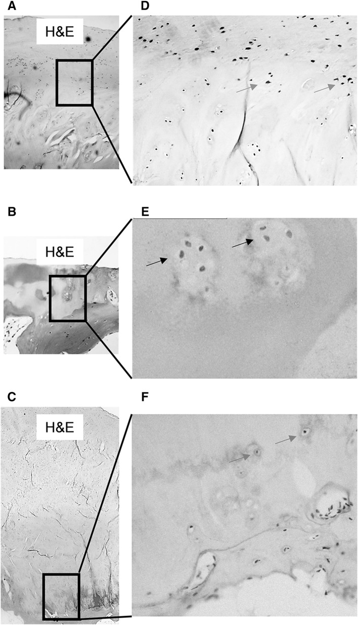
Fig. 2.
DKK-1 expression is high in OA chondrocytes Cylindrical cores were taken from OA femoral head biopsies, fixed in paraformaldehyde, decalcified in EDTA, 5 µm serial sections were cut longitudinally and stained with anti-human DKK-1 antibody. a–c Representative H&E staining of cartilage in cores taken from macroscopically normal cartilage, partial cartilage, and osteophyte regions. DKK-1 immunostaining only observed in chondrocytes in cores taken from macroscopically normal cartilage (blue arrows in d), but not in partial cartilage defect (black arrows in e) or osteophyte regions (red arrows in f) respectively. a–c ×4, d and f ×10 and e ×20 magnification

