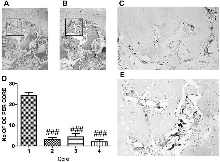
Fig. 6.
Osteoclast number is high in OA macroscopically normal cartilage Cylindrical cores were taken from OA femoral head biopsies, fixed in paraformaldehyde, decalcified in EDTA, 5 µm serial sections were cut longitudinally, and stained with anti-human CATK antibody. a–c Representative staining of serial sections of cartilage in core taken from full thickness cartilage. a H&E staining, b IgG negative staining, c CATK immunostaining, d quantification of CATK positive osteoclasts (OC) in cores from full thickness cartilage (1), partial cartilage (2), full cartilage defect (3) and osteophyte (4). e High magnification of immunoreactive osteoclasts from slide c. a and b ×4, c ×10 and e ×20 magnification. Data presented as mean ± SEM (One-way analysis of variance, Tukey’s multiple comparison test) of positive osteoclasts from 6 slides per each core from 4 femoral head biopsies. ### P < 0.001 for comparison of immunoreactive osteoclasts from cores relative to core 1

